
Anatomy
The stifle is the equivalent of the human knee, and it is the largest, most complex joint in the horse. The bones that make up the stifle are the femur (thigh), tibia (shin), and patella (kneecap). The stifle lifts the leg upward and forward, so it's pretty critical to moving, never mind athletic pursuits. A thin capsule surrounds the entire stifle joint that has a specialized fluid to help with shock absorption and lubrication. There are lots of ligaments -- that is, fibrous connective tissue which attaches bone to bone -- that hold things together and keep the joint stable. Here are the key ligaments (click to enlarge the image):
- The medial and lateral patellar ligaments attach the patella to the tibia.
- Short, thick collateral ligaments located on the inside and outside of the stifle joint attach the femur and the tibia. They are not shown on the diagram to the right but can be seen here.
- The medial meniscus and lateral meniscus are semicircular pads of thick, resilient fibrocartilage. They occupy the spaces between the ends of the femur and the tibia. These pads help cushion the weight-bearing forces in the stifle.
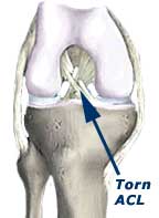
- Two strong ligamentous bands, the anterior (forward) and posterior (hind) cruciate ligaments, crisscross within the joint and span between attachment sites on the femur and the tibia. You can see them in the caudal diagram link below. Cruciate ligaments provide support for the joint when it is pushed excessively forward or backward. These ligaments also provide some rotational stability.

 What can go wrong
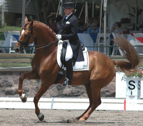
Many vets feel that stifle injuries are underdiagnosed, in part because the stifle is difficult to evaluate. The stifle joint is subject to the same kinds of troubles that other joints/bones might have: OCD, bone cysts, physitis (swelling at the growth plates), arthritis, etc. Traumatic injury to the bones and ligaments are also possible. Activities that involve sharp turns while "digging in the ground" are likely to result in stifle trauma -- in dressage, the pirouette and tempi changes stress the stifle. Cutting and barrel horses tend to have stifle problems as well. Jumping horses can strain the meniscal ligaments. Subloxation of the patella, which locks the stifle in an extended position, is a common problem with horses, especially straight-hocked horses -- but I won't discuss it in depth here.
 Ligament damage
Different ligaments are diagnosed and handled differently. Horses tend to tear meniscal ligaments. Meniscal injuries are found in 10% of all stifle lameness cases. Usually there is swelling and overt lameness. Arthroscopy is the best way to diagnose meniscal injuries, but MRI is becoming more available. Treatment of meniscal damage relies on extended rest, and if you have the money, and injection with Interleukin-1 receptor antagonist protein (IRAP) or stem cells. A meniscal tear should be non-weight-bearing for 60 days, and this is clearly not possible with horses. Surgery is not usually very successful.
Cruciate ligaments connect the femur to the tibia, and they prevent excessive bending--tears can lead to joint instability. They are easily damaged. Lameness severity tends to mirror the seriousness of the tear. Prognosis for complete tears is very guarded, and even if the tear is partial, only about 50% of the injured animals return to their former level of work.
With a collateral ligament injury,lameness may be progressive over several months, and there may be concurrent injury. Diagnostic ultrasound and bone scans are the best tools to identify this problem. These injuries need rest and a slow rehabilitation. Despite careful rehab, most horses have their level of activity limited by this injury. Stem cell therapy might be something to try.
Symptoms
Horses with a stifle issue may tend to stand with stifle rotated out. They may put weight on toe while standing or moving, and sometimes the hoof will show excessive wear on the toe. Stifle pain makes the horse reluctant to reach forward with the hind leg, making the stride short and stabby, landing on the toe. The foot flight will stay low to the ground to reduce limb flexion. This causes a pronounced hip hike. Lameness often is more evident when the affected limb is on the outside of the circle. The horse may "bunny hop" annd try to canter when asked to move forward. The horse may be reluctant to go down an incline.
Diagnosis
There are several techniques vets use for diagnosing stifle issues...
- A hands-on exam is used to feel effusion, ridges, spurs, and ligament thickening.
- Anesthesia/blocking of the joints to isolate the source of lameness.
- Radiographs are often done to detect bony changes, but they can be inconclusive since there are several soft tissue structures in this joint that will not show in an x-ray.
- Ultrasound gives some good images of these structures but is also limited by (depth x resolution. Also and also due to anatomical structures may be positioned in unaccessible places. Ultrasound is effective in diagnosing collateral ligament injuries
- Magnetic resonance imaging (MRI) is capable of imaging the stifle provided a horse's leg can be fully loaded into the machine.

- Thermography isn't mentioned a lot as a diagnostic technique, but look at the the picture to the right depicting stifle issues -- downright pretty!
- Nuclear scintigraphy (bone scan) is sometimes used but results are spotty --
- Computed tomography (CT) might be able to image the stifle with detail approaching that of MRI, and with a shorter examination time. (The Horse, 2008)
- Arthroscopy is the ultimate diagnostic procedure, providing the best visualization of the joint interior.

Prognosis
The prognosis depends on the severity of the condition/injury, how quickly it is detected, and the form of treatment used. The longer a condition goes untreated, the more likely secondary damage (arthritis, degenerative joint disease, etc.) will occur. Unfortunately, from what I've read, most adult horses do not return to their former level of work.
RESOURCES
Don't trifle with the stifle from The Horse magazine
Lameness may hinge on the stifle, Thoroughbred Times, Feb 27,2006
Diagnosis and Treatment of Stifle Lameness from The Horse magazine
VMED 5362 Diseases of the Horse Stifle Lameness from Louisiana State University
Lameness of the Horse by John Victor LaCroix (stifle section)
Strained stifle in horses from Equisearch
Dealing with stifle injuries from Horse and Hound
Chapter on the Stifle from Adams lameness in horses
Inside the equine stifle from Veterinary News
Treating stifle injuries from The Horse magazine
Treatment of a stifle strain from Equisearch
Stifle lameness in the athletic horse from AAEP Focus
Horse anatomy: The stifle
Lameness associated with the stifle and pelvic regions from AAEP 2002 Proceedings
Diagnosing stifle disease from The Horse magazine
Problem with stifling from horseinformation.com
Section of the stifle of Horse Conformation by Equine Research


.jpg)


Thanks so much on your article, it had a lot of good information. I ride hunters and equitation but started my riding in dressage. The stifle is a very important piece to the horse and it was good to get some more info on it.
ReplyDeleteGreat article - keep up the good work :)
ReplyDeleteAnother thank you for your article, it's well written and explanatory. My horse has stifle problems but his vet failed to diagnose it during a rather weak lameness exam. Research of my own is pointing to a collapsing stifle with perhaps cruciate ligament trouble. Is it ok to reference and link this article on my blog?
ReplyDeleteYes, thanks, I'm flattered! I'll check out your entry...
ReplyDeleteI've been reading this a lot lately with Gus's stifle injury. Thank you!
ReplyDeleteThanks for this summary. I've just had my horse diagnosed with a stifle problem through nerve blocks. I've now got to decide what next.. do I go for a steroid injection or arthroscope so reading up as much as I can!
ReplyDeleteHow has Dauphin's recovery gone? Is he back to work? At what level was he able to return to work? Stifle injuries are so slow and hard to rehab, but patience is the key. Hopefully Dauphin has recovered and is back to work. It'd be nice to hear how he is doing. Please keep us updated. Thank you for your post.
ReplyDeletequarter horse information
ReplyDelete